What is Vascular Ultrasound?
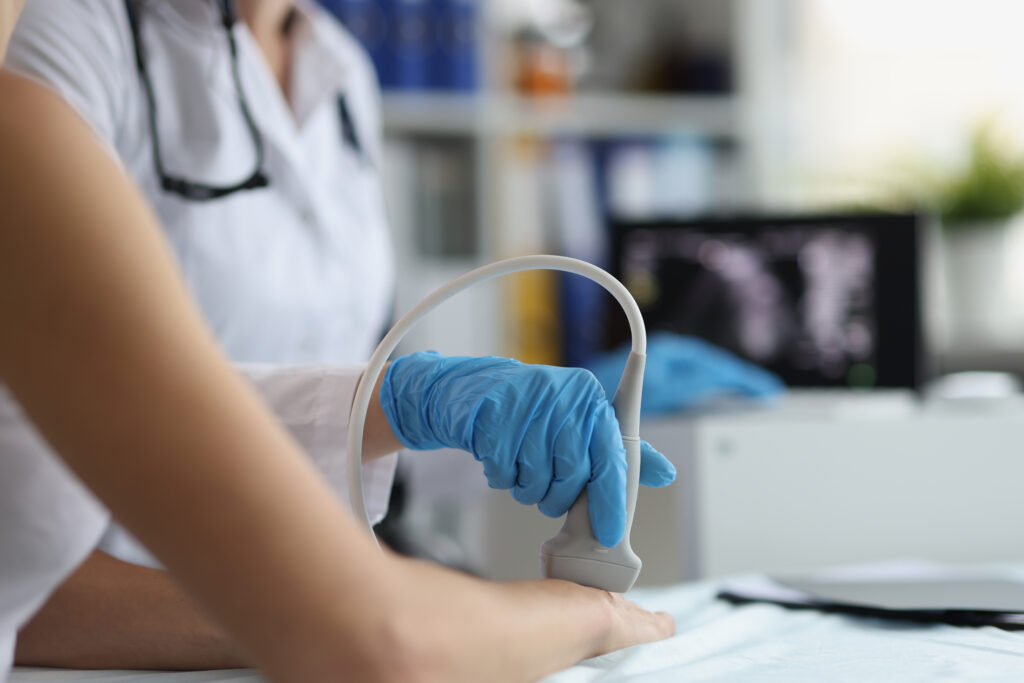
Vascular ultrasound, also known as a duplex study, is a non-invasive diagnostic imaging procedure that uses high-frequency sound waves to evaluate the circulatory system, including the veins and arteries. This technology provides detailed images that allow healthcare professionals to assess blood flow and detect blockages or abnormalities in the vessels.
Why is Vascular Ultrasound Important?
Vascular ultrasound is crucial in diagnosing and monitoring various vascular conditions, including:
- Atherosclerosis: This condition involves the narrowing of arteries due to plaque buildup, which can lead to cardiovascular complications.
- Deep Vein Thrombosis (DVT): A blood clot formation in a deep vein, usually in the legs, can cause serious health risks if left untreated.
- Peripheral Artery Disease (PAD): Reduced blood flow to the limbs, leading to pain and potential tissue damage.
- Aneurysms: Abnormal bulges in the walls of arteries can pose life-threatening risks if they rupture.
- Varicose Veins: Enlarged veins often cause discomfort and cosmetic concerns.
With vascular ultrasound, healthcare providers can quickly diagnose these conditions and monitor ongoing treatments’ effectiveness, ensuring optimal patient care and outcomes.
Preparation for the Procedure
Preparing for a vascular ultrasound is straightforward and generally requires minimal effort from the patient:
- Clothing: Wear comfortable, loose-fitting clothing. You may be asked to wear a gown during the procedure.
- Fasting: Typically, no fasting is required, but your doctor may provide specific instructions depending on the area being examined.
- Medications: Continue to take prescribed medications unless instructed otherwise by your healthcare provider.
- Personal Items: Remove any jewelry or accessories that may interfere with the ultrasound process.
What to Expect During the Ultrasound
The vascular ultrasound procedure is safe, painless, and usually takes about 30 to 60 minutes, depending on the specific area being examined. Here’s what you can expect:
- Positioning: You will lie on an examination table, and a trained sonographer will apply a warm, water-based gel to the skin over the area to be examined. This gel helps the ultrasound transducer glide smoothly and improve sound wave transmission.
- Imaging: The sonographer will move the transducer over the skin, capturing real-time images of your blood vessels. You may hear pulse-like sounds during the examination as the machine records blood flow.
- Comfort: You may be asked to change positions or hold your breath for a short time to obtain clearer images. Rest assured, the sonographer will ensure you are comfortable throughout the process.
- Post-Procedure: After the procedure, the gel will be wiped off your skin, and you can resume normal activities immediately. There are no known risks associated with vascular ultrasound, making it a safe option for patients of all ages.
Understanding Your Results
After the examination, the images are analyzed by a radiologist or vascular specialist who will provide a detailed report to your referring healthcare provider. This report will help determine the best course of action, whether lifestyle modifications, medications, or further interventions are needed.
Why Choose Our Medical Imaging Center?
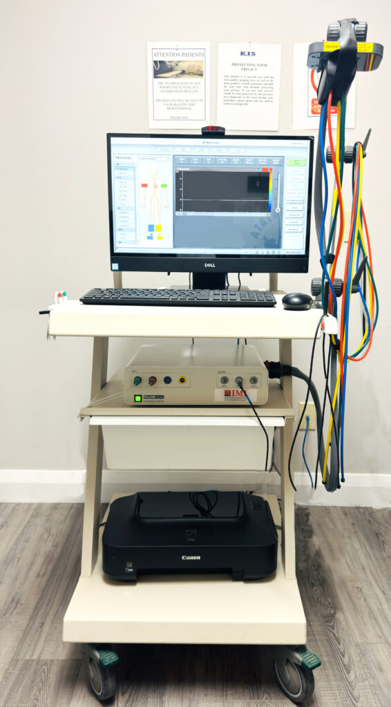
At KIS, we are committed to delivering top-quality care and advanced diagnostic services. Our state-of-the-art ultrasound technology, combined with our experienced team of professionals, ensures accurate and timely results. We prioritize patient comfort and satisfaction, offering a seamless experience from scheduling to follow-up care.
For more information or to schedule your vascular ultrasound appointment, please contact us today. We are here to assist you in your healthcare journey and provide the exceptional care you deserve.

